What is a Brain MRI?
A Brain MRI (Magnetic Resonance Imaging) is a non-invasive diagnostic imaging procedure that uses powerful magnets and radio waves to create detailed images of the brain and its structures. Unlike X-rays or CT scans, Brain MRIs do not use ionizing radiation, making them a safer option for repeated imaging. This advanced imaging technique provides high-resolution images that help healthcare providers diagnose and monitor a wide range of neurological conditions, including tumors, strokes, multiple sclerosis, and traumatic brain injuries. The procedure is painless and typically takes between 30 to 60 minutes, depending on the specific requirements of the examination.
Who Can Take the Brain MRI?
A Brain MRI is recommended for individuals who:
- Are Experiencing Neurological Symptoms: Such as severe headaches, dizziness, seizures, memory loss, or changes in vision.
- Have a History of Brain Disorders: Including epilepsy, multiple sclerosis, or previous brain injuries.
- Are Suspected of Having a Brain Tumor: To detect and monitor the size and location of tumors.
- Have Undiagnosed Neurological Conditions: When other diagnostic tests are inconclusive.
- Are Preparing for Brain Surgery: To provide detailed images for surgical planning.
- Have Suffered a Stroke: To assess the extent of brain damage and guide treatment.
- Are Pregnant: When necessary, as MRI does not use ionizing radiation, though it's typically avoided during the first trimester unless essential.
- Have a Family History of Neurological Disorders: Increasing the risk of hereditary brain conditions.
- Are Undergoing Treatment for Brain Conditions: To monitor the effectiveness of therapies and track disease progression.
- Have Persistent or Recurrent Infections: Such as meningitis or encephalitis, affecting the brain and its surrounding structures.
- Have Cognitive or Behavioral Changes: That may indicate underlying brain abnormalities.
- Are Athletes: Who have sustained concussions or other head injuries and require detailed brain imaging.
- Have Undocumented Medical Records: To establish a comprehensive baseline for future comparisons and evaluations.
When Can the Brain MRI Be Performed?
The timing for a Brain MRI depends on various factors, including the onset of symptoms, medical history, and specific health concerns:
- When Symptoms Arise: Such as sudden or severe headaches, seizures, or neurological deficits.
- During Routine Health Check-ups: For individuals with risk factors for brain disorders.
- Before and After Brain Surgeries: To assess surgical outcomes and monitor recovery.
- When Diagnosing Specific Conditions: Such as brain tumors, multiple sclerosis, or traumatic brain injuries.
- After a Head Injury: To evaluate potential brain damage or bleeding.
- When Other Diagnostic Tests are Inconclusive: To obtain more detailed images and a more accurate diagnosis.
- For Research Purposes: To study brain structures and functions in various neurological conditions.
- During Pregnancy: When necessary, and after consulting with a healthcare provider.
- For Monitoring Chronic Conditions: Like epilepsy or neurodegenerative diseases, to manage and adjust treatment plans effectively.
- When Evaluating Cognitive Decline: In older adults to differentiate between types of dementia.
- For Pediatric Assessments: To diagnose and monitor brain development issues or congenital anomalies.
- In Emergency Situations: Such as suspected stroke or acute neurological events, to guide immediate treatment.
Procedure and Duration
The Brain MRI procedure is straightforward, non-invasive, and typically completed within a short timeframe:
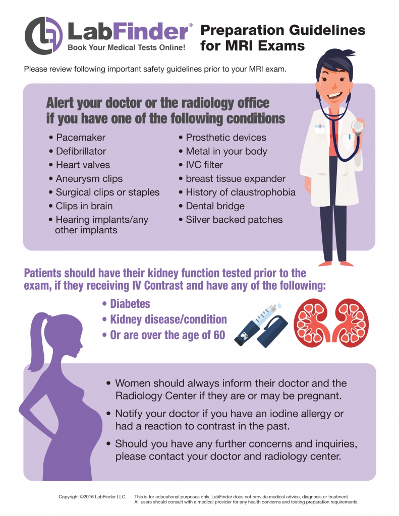
- Preparation: No special preparation is usually required. However, inform your healthcare provider about any implants, pacemakers, or metal devices, as these can interfere with the MRI. You may also be asked to remove jewelry, watches, and other metal objects before the scan.
- Positioning: You will lie down on a motorized table that slides into the MRI machine, which is a large, tube-shaped device. Cushions and straps may be used to help you remain still during the procedure.
- The Scan: The MRI machine uses a strong magnetic field and radio waves to generate detailed images of the brain. You may hear loud tapping or thumping noises during the scan, and earplugs or headphones are usually provided to reduce discomfort.
- Duration: The entire procedure typically takes between 30 to 60 minutes, depending on the complexity of the examination and the specific requirements.
- Post-Scan: You can resume normal activities immediately after the scan. There are no restrictions unless advised by your healthcare provider.
Related Conditions or Illnesses
A Brain MRI helps diagnose and monitor several neurological conditions, including:
- Brain Tumors: Identifying the presence, size, and location of tumors.
- Stroke: Assessing the extent of brain damage caused by interrupted blood flow.
- Multiple Sclerosis (MS): Detecting lesions and monitoring disease progression.
- Traumatic Brain Injury (TBI): Evaluating structural damage from head injuries.
- Epilepsy: Identifying structural abnormalities that may cause seizures.
- Parkinson’s Disease: Monitoring changes in brain structures associated with the disease.
- Alzheimer’s Disease and Other Dementias: Assessing brain atrophy and other changes related to cognitive decline.
- Hydrocephalus: Detecting excess cerebrospinal fluid in the brain.
- Infections: Such as meningitis or encephalitis, affecting the brain and its surrounding structures.
- Developmental Disorders: In children, identifying congenital anomalies or developmental delays.
- Neurodegenerative Diseases: Like Huntington’s disease, assessing brain structure and function.
- Vascular Malformations: Detecting abnormal blood vessel formations in the brain.
- Chiari Malformations: Identifying structural defects in the cerebellum.
- Epileptic Foci: Locating specific areas in the brain responsible for seizure activity.
- Brain Infections: Such as abscesses or encephalitis, assessing their location and extent.
- Brain Inflammation: Detecting areas of inflammation that may be indicative of various conditions.
- Sleep Disorders: Investigating structural causes of sleep apnea or other sleep-related issues.
- Mood Disorders: Exploring structural and functional brain changes associated with depression, bipolar disorder, and anxiety.
- Autism Spectrum Disorders: Researching brain structure and connectivity in individuals with autism.
Risks
While a Brain MRI is considered very safe, there are some potential risks and considerations:
- Metal Implants and Devices: The strong magnetic field can interact with certain implants, pacemakers, cochlear implants, or metal fragments in the body, posing safety risks.
- Claustrophobia: The enclosed space of the MRI machine can cause anxiety or panic in individuals with claustrophobia. Open MRI machines or sedation options may be available to alleviate discomfort.
- Contrast Dye Reactions: In some cases, a contrast agent (gadolinium-based) may be injected to enhance image quality. Rarely, individuals may experience allergic reactions, including rash, itching, or more severe reactions like anaphylaxis.
- Heating of Metallic Objects: Metal jewelry or accessories can heat up during the scan, causing burns or discomfort.
- Noise-Induced Hearing Damage: The loud noises produced by the MRI machine can potentially damage hearing if proper ear protection is not used.
- False Positives/Negatives: Inaccurate interpretations can occur due to overlapping structures, motion artifacts, or technical errors, potentially leading to unnecessary further testing or missed diagnoses.
- Kidney Function Concerns: Individuals with severe kidney disease may be at risk for a rare condition called nephrogenic systemic fibrosis (NSF) when exposed to certain gadolinium-based contrast agents.
- Pregnancy Considerations: While MRI is generally considered safe during pregnancy, especially after the first trimester, the use of contrast agents is typically avoided unless absolutely necessary.
- Emotional Impact: Discovering abnormalities can cause anxiety or stress, even if they turn out to be benign.
Preparations
Preparing for a Brain MRI involves several important steps to ensure accurate results and a comfortable experience:
- Inform Your Provider of Implants and Medical Devices: Disclose any metal implants, pacemakers, cochlear implants, or other medical devices that could be affected by the MRI's magnetic field.
- Avoid Wearing Metal Objects: Remove jewelry, watches, glasses, and any metal accessories before the scan.
- Follow Fasting Instructions: If a contrast agent is to be used, you may be instructed to fast for a few hours before the test.
- Wear Comfortable Clothing: Opt for loose-fitting clothes without metal fasteners or buttons. A gown may be provided by the imaging center.
- Arrange Transportation if Sedation is Required: If you are to receive sedation to alleviate claustrophobia, ensure you have someone to drive you home after the procedure.
- Stay Hydrated: Drink plenty of water before the test unless instructed otherwise by your healthcare provider.
- Communicate with Your Provider: Discuss any concerns or questions you have about the MRI to ensure you are fully prepared.
- Bring Previous Imaging Results: If you have had prior MRIs or other brain imaging tests, bring the results for comparison.
- Prepare for the Noise: Earplugs or headphones are usually provided to reduce the loud noises produced by the MRI machine.
- Stay Relaxed: Try to remain calm and still during the procedure to ensure clear and accurate images.
- Follow Specific Instructions: Adhere to any additional guidelines provided by your healthcare provider or the imaging center.

Other Similar Tests
There are several other tests related to diagnosing and assessing brain conditions and overall neurological health:
- CT Scan (Computed Tomography): Uses X-rays to create detailed cross-sectional images of the brain, useful for detecting acute injuries, hemorrhages, and tumors.
- PET Scan (Positron Emission Tomography): Assesses brain function and metabolism by detecting radioactive tracers, useful in diagnosing cancer, Alzheimer's disease, and other neurological conditions.
- fMRI (Functional Magnetic Resonance Imaging): Measures and maps brain activity by detecting changes in blood flow, used in research and for planning brain surgery.
- EEG (Electroencephalogram): Records electrical activity in the brain, useful for diagnosing epilepsy and other seizure disorders.
- MEG (Magnetoencephalography): Maps brain activity by recording magnetic fields produced by neural activity, used in research and clinical settings.
- Angiography: Visualizes blood vessels in the brain using contrast agents and X-ray imaging, useful for detecting aneurysms, stenosis, and vascular malformations.
- Neuropsychological Testing: Assesses cognitive functions, memory, problem-solving, and other mental processes to diagnose conditions like dementia or brain injuries.
- Spinal Tap (Lumbar Puncture): Collects cerebrospinal fluid for analysis to diagnose infections, hemorrhages, or multiple sclerosis.
- Single-Photon Emission Computed Tomography (SPECT): Similar to PET scans, it evaluates blood flow and activity in the brain.
- Diffusion Tensor Imaging (DTI): A type of MRI that maps the diffusion of water in brain tissue, useful for visualizing white matter tracts.
- Transcranial Doppler Ultrasound: Measures blood flow velocity in the brain's blood vessels, used to detect vasospasm or other vascular conditions.
- Magnetic Resonance Spectroscopy (MRS): An MRI-based technique that measures the chemical composition of brain tissue.
- Biopsy: In rare cases, a tissue sample from the brain may be taken for laboratory analysis to diagnose tumors or other conditions.
- Ultrasound: Used primarily in infants to image the brain through the fontanelles, as the skull bones are not fully fused.
- Oculomotor Testing: Evaluates eye movements to assess brain function and detect neurological abnormalities.
- Vestibular Testing: Assesses balance and spatial orientation, useful in diagnosing vestibular disorders.
How Accurate is the Brain MRI?
A Brain MRI is highly accurate in diagnosing a wide range of neurological conditions when performed correctly. The use of strong magnetic fields and advanced imaging technology allows for the detailed visualization of brain structures, making it effective in detecting abnormalities such as tumors, lesions, vascular malformations, and areas of inflammation. The accuracy depends on several factors, including the quality of the MRI machine, the skill of the technician, proper patient positioning, and the specific imaging protocols used. High-resolution MRI scanners provide superior image clarity, enhancing the detection of even small or subtle changes in brain anatomy. However, certain factors like patient movement, metallic implants, or the presence of hair can affect image quality. When combined with other diagnostic tests and clinical evaluations, Brain MRI offers a comprehensive assessment of brain health. It is essential to have the MRI interpreted by a qualified radiologist or neurologist to ensure accurate diagnosis and appropriate treatment planning.
What Should I Do If I Find Something Concerning on a Brain MRI?
If your Brain MRI results indicate any abnormalities, here's what you should do next:
- Consult Your Neurologist or Healthcare Provider: Discuss the findings in detail to understand their implications and determine the necessary next steps.
- Schedule Follow-Up Tests: Additional diagnostic procedures, such as PET scans, EEGs, or biopsies, may be required to confirm and further investigate the findings.
- Consider Specialist Referrals: Depending on the abnormality, you may need to consult with an oncologist, neurosurgeon, or other specialists for further evaluation and treatment.
- Develop a Treatment Plan: Work with your healthcare provider to create a plan to address the identified condition, which may include medications, surgery, radiation therapy, or other interventions.
- Adhere to Medical Recommendations: Follow prescribed treatments and recommendations strictly to ensure effective management of the condition.
- Implement Preventive Measures: Adopt recommended lifestyle changes, such as improving diet, increasing physical activity, managing stress, and avoiding harmful substances to support overall brain health.
- Monitor Symptoms: Keep track of any new or worsening symptoms and report them to your healthcare provider promptly.
- Seek Support: Reach out to support groups, counseling services, or trusted individuals if you're dealing with significant health changes or emotional stress related to the findings.
- Maintain Regular Check-Ups: Schedule and attend regular medical appointments to monitor your condition and adjust treatment plans as necessary.
- Address Underlying Causes: Work with your healthcare provider to identify and treat any underlying conditions contributing to abnormal MRI results.
- Educate Yourself: Learn about the diagnosed condition, its management, and preventive measures to make informed decisions about your health.
- Follow Preventive Measures: If the MRI detects a condition that can be managed or prevented, adhere to your healthcare provider's recommendations to maintain your health.
- Stay Informed on Treatment Advances: Keep up-to-date with the latest treatments and recommendations for your condition by consulting reputable medical sources or your healthcare provider.
Book Brain MRI Using LabFinder
Booking your Brain MRI is now easier than ever with LabFinder. LabFinder allows you to locate participating labs and imaging centers near you, ensuring prompt and reliable service. Many of these facilities accept insurance, making the process hassle-free. So, if you're looking for a "brain mri near me," or "brain mri scan near me," you've come to the right place. Schedule your Brain MRI online and save time by avoiding long waits or multiple phone calls.
Conclusion
A Brain MRI is an essential diagnostic tool for assessing and managing neurological health. By providing detailed and accurate images of the brain and its structures, it aids in the early detection and precise diagnosis of a wide range of conditions, from tumors and strokes to multiple sclerosis and traumatic injuries. Understanding what the test entails, who should take it, and the procedures involved empowers you to make informed decisions about your neurological health and take proactive steps toward preventing and managing brain-related conditions. Whether you're experiencing symptoms, undergoing routine screenings, or preparing for brain surgery, a Brain MRI offers the clarity and precision needed for effective diagnosis and successful treatment outcomes. Don’t wait—book your Brain MRI near you with LabFinder today and take proactive steps toward maintaining your brain and overall health journey.
Book on LabFinder: find a lab today on our lab finder and request a test doctor guided.

