What is an Orthopantogram (Panoramic Dental X-ray)?
An Orthopantogram, commonly known as a Panoramic Dental X-ray, is a comprehensive imaging technique that captures a wide-view, two-dimensional image of the entire mouth, including the teeth, jaws, nasal area, and surrounding structures. Unlike traditional intraoral X-rays that focus on specific areas, a panoramic X-ray provides a full overview of the oral and maxillofacial region, making it an invaluable tool for dentists and oral surgeons in diagnosing and planning various dental treatments. This non-invasive procedure utilizes low-dose radiation to produce detailed images, aiding in the detection of dental issues that may not be visible through regular examinations.
Who Can Take the Orthopantogram (Panoramic Dental X-ray)?
An Orthopantogram is recommended for individuals who:
- Are Planning Dental Implants: To assess bone structure and density for optimal implant placement.
- Have Impacted Wisdom Teeth: To determine the position and potential complications of wisdom teeth before extraction.
- Require Orthodontic Treatment: To evaluate jaw alignment and plan effective orthodontic interventions.
- Have Persistent Dental Pain or Infections: To identify underlying issues such as abscesses or cysts that may not be visible during a standard dental exam.
- Are Undergoing Oral Surgery: To provide detailed imaging for surgical planning and to minimize risks.
- Have a History of Jaw Fractures or Abnormalities: To monitor bone health and structural integrity.
- Are Experiencing TMJ Disorders: To assess the temporomandibular joint and surrounding structures for abnormalities.
- Need Evaluation of Bone Density: To detect bone loss or other skeletal issues that can affect dental health.
- Have Undocumented Dental Records: To establish a comprehensive baseline for future treatments and comparisons.
- Are Preparing for Dental Prosthetics: Such as dentures or bridges, to ensure proper fit and alignment.
- Are High-Risk Individuals: With conditions that may affect dental or jaw health, necessitating detailed imaging.
When Can the Orthopantogram (Panoramic Dental X-ray) Be Performed?
The timing for an Orthopantogram depends on various factors, including dental needs, symptoms, and treatment plans:
- Before Dental Implant Placement: To ensure sufficient bone structure and optimal positioning.
- During Orthodontic Evaluation: To assess jaw alignment and plan braces or aligner treatments effectively.
- When Diagnosing Impacted Teeth: Such as wisdom teeth, to determine their position and potential complications.
- For Preoperative Planning: Prior to oral surgeries to map out the surgical approach and minimize risks.
- When Experiencing Persistent Dental Pain: To identify hidden infections, cysts, or abscesses causing discomfort.
- During Routine Dental Check-ups: Especially for individuals with risk factors for dental or jaw conditions.
- After Jaw Injuries: To evaluate bone healing and detect any structural issues post-injury.
- When Monitoring Chronic Conditions: Such as osteoporosis, which can affect bone density and dental health.
- For Evaluation of TMJ Disorders: To assess the temporomandibular joint and surrounding structures.
- Prior to Dental Prosthetic Fitting: To ensure accurate fitting and alignment of dentures, bridges, or other prosthetics.
- When Other Imaging Tests are Inconclusive: To obtain a more comprehensive view of the oral and maxillofacial region.
Procedure and Duration
The Orthopantogram (Panoramic Dental X-ray) procedure is quick, painless, and typically completed within a short timeframe:
- Preparation: Wear comfortable clothing and remove any metal jewelry or accessories that might interfere with the imaging.
- Positioning: You will be seated in a dental chair with your head positioned against the X-ray machine's receptor. The technician will guide you to remain still while aligning your head and jaw properly.
- The Scan: The X-ray machine rotates around your head, capturing multiple images from different angles to create a comprehensive panoramic view.
- Duration: The entire procedure typically takes between 10 to 20 minutes, depending on the complexity of the scan and the specific requirements.
- Post-Scan: You can resume normal activities immediately after the scan. There are no restrictions unless advised by your healthcare provider.
Related Conditions or Illnesses
An Orthopantogram helps diagnose and monitor several dental and oral conditions, including:
- Dental Caries (Cavities): Identifies areas of tooth decay that may not be visible during a standard dental exam.
- Impact of Wisdom Teeth: Determines if wisdom teeth are impacted, their position, and potential complications.
- Jaw Fractures: Detects fractures or structural abnormalities in the jawbone.
- Bone Loss: Assesses bone density around teeth, which can indicate periodontal disease or osteoporosis.
- Oral Cysts and Tumors: Identifies benign or malignant growths within the oral cavity.
- TMJ Disorders: Evaluates the temporomandibular joint for abnormalities or structural issues.
- Dental Implants Planning: Maps out bone structure and density to ensure successful implant placement.
- Orthodontic Planning: Assists in designing effective braces or aligner treatments by providing a comprehensive view of the jaw and teeth alignment.
- Prosthetic Fitting: Ensures accurate fitting of dentures, bridges, or other dental prosthetics by assessing jaw and bone structure.
- Sinus Evaluation: Detects sinus infections or abnormalities that may affect dental health.
- Root Canal Treatment: Identifies the extent of root damage or infection requiring endodontic procedures.
- Oral Infections: Detects abscesses or other infections within the oral cavity.
- Developmental Abnormalities: Identifies congenital defects affecting the oral and maxillofacial region.
- Dental Trauma: Assesses the extent of injuries to teeth and jaw following accidents or trauma.
- Sinusitis: Evaluates inflammation or infection in the sinus cavities that can impact dental health.
Risks
The Orthopantogram (Panoramic Dental X-ray) is considered safe, with minimal risks involved:
- Radiation Exposure: Involves a low dose of ionizing radiation, which is minimal compared to the diagnostic benefits. Protective measures, such as lead aprons, are used to shield parts of the body not being imaged.
- Discomfort: Positioning during the scan may cause temporary discomfort, especially for individuals with limited mobility or pain from dental issues.
- Allergic Reactions: Extremely rare, unless a contrast agent is used, which is uncommon for standard panoramic X-rays.
- False Results: Inaccurate interpretations can occur due to overlapping structures, previous dental surgeries, or poor image quality, potentially leading to unnecessary further testing or missed diagnoses.
- Emotional Impact: Discovering abnormalities can cause anxiety or stress, even if they turn out to be benign.
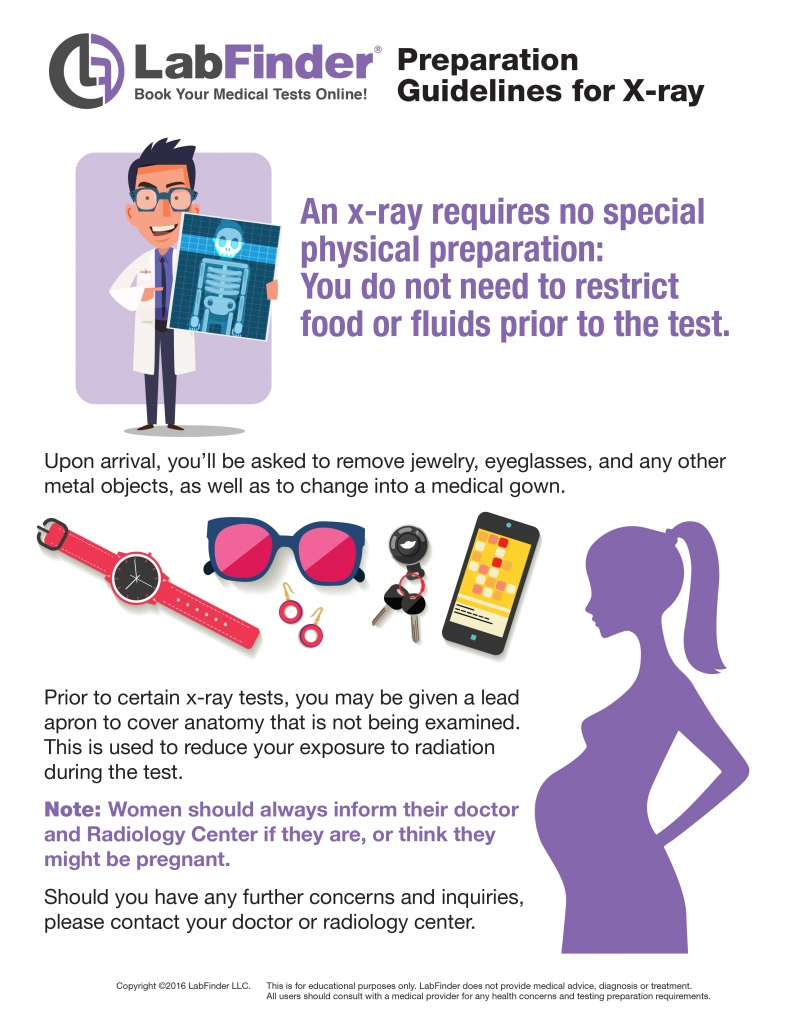
Preparations
Preparing for an Orthopantogram involves a few simple steps to ensure accurate results and a comfortable experience:
- Wear Comfortable Clothing: Choose clothing without metal buttons, zippers, or accessories that could interfere with the imaging.
- Remove Metal Objects: Take off jewelry, watches, and other metal items before the scan.
- Inform Your Provider: Let your dentist know if you have any dental implants, braces, or other metal appliances that may affect the scan.
- Avoid Eating or Drinking Immediately Before the Test: Especially if the scan is scheduled early in the morning.
- Follow Specific Instructions: Adhere to any additional guidelines provided by your healthcare provider or the imaging center.
- Stay Relaxed and Still: Try to remain calm and avoid movement during the procedure to ensure clear and accurate images.
- Bring Previous Imaging Results: If you have had prior X-rays or imaging tests, bring the results for comparison.
Other Similar Tests
There are several other dental imaging tests related to Orthopantograms, each with its specific uses:
- Intraoral X-rays: Includes bitewing and periapical X-rays for detailed images of specific teeth and surrounding bone structures.
- Cephalometric X-ray: Primarily used in orthodontics to assess the relationships between dental and skeletal structures.
- Dental CBCT (Cone Beam Computed Tomography): Provides three-dimensional imaging for more detailed assessments compared to panoramic X-rays.
- Panoramic Tomosynthesis: An advanced form of panoramic imaging that reduces image overlap and improves diagnostic accuracy.
- Digital Panoramic X-ray: A modern version of the panoramic X-ray that offers digital image storage and enhanced image quality.
- Bone Scans: Detect abnormalities in bone metabolism and detect tumors or fractures.
- MRI (Magnetic Resonance Imaging): Provides detailed images of soft tissues without radiation, useful for complex dental and jaw issues.
- Ultrasound: Used for evaluating soft tissue structures in the oral and maxillofacial region.
- Optical Coherence Tomography (OCT): A non-invasive imaging test that uses light waves to take cross-section pictures of tissues.
- Fluorescence Imaging: Utilizes special light to detect early signs of tooth decay and other oral conditions.
How Accurate is the Orthopantogram (Panoramic Dental X-ray)?
An Orthopantogram (Panoramic Dental X-ray) is highly accurate in providing a comprehensive overview of the dental and maxillofacial structures. The combination of multiple angles captured during the scan allows for the detection of a wide range of conditions, from tooth decay and impacted wisdom teeth to bone fractures and tumors. The accuracy depends on factors such as the quality of the imaging equipment, the skill of the technician, and proper patient positioning. Modern digital panoramic X-rays offer enhanced image clarity and reduced radiation exposure compared to traditional film-based systems. However, certain factors like excessive dental restorations, metal implants, or patient movement can affect image quality and diagnostic accuracy. It is essential to have the X-ray interpreted by a qualified radiologist or dental specialist to ensure accurate diagnosis and appropriate treatment planning.
What Should I Do If I Find Something Concerning on a Orthopantogram (Panoramic Dental X-ray)?
If your Orthopantogram results indicate any abnormalities, here's what you should do next:
- Consult Your Dentist or Oral Surgeon: Discuss the findings in detail to understand their implications and determine the necessary next steps.
- Schedule Follow-Up Tests: Additional imaging or diagnostic procedures, such as CBCT scans, MRIs, or biopsies, may be required to confirm and further investigate the findings.
- Consider Specialist Referrals: Depending on the abnormality, you may need to consult with an orthodontist, oral surgeon, oncologist, or other specialists for further evaluation and treatment.
- Develop a Treatment Plan: Work with your healthcare provider to create a plan to address the identified condition, which may include medications, surgical interventions, orthodontic treatments, or other procedures.
- Stay Informed: Educate yourself about the condition and potential treatments to make informed decisions about your dental health.
- Seek Support: Reach out to support groups, counseling services, or trusted individuals if you're dealing with significant health changes or emotional stress related to the findings.
- Follow Preventive Measures: If the X-ray detects a condition that can be managed or prevented, adhere to your healthcare provider's recommendations to maintain your health.
- Maintain Regular Check-Ups: Schedule and attend regular dental appointments to monitor your condition and adjust treatment plans as necessary.
- Address Underlying Causes: Work with your healthcare provider to identify and treat any underlying causes contributing to abnormal Orthopantogram results.
- Consider Lifestyle Modifications: Implement recommended lifestyle changes, such as improving oral hygiene, diet adjustments, or quitting smoking, to support overall dental health.
Book Orthopantogram (Panoramic Dental X-ray) Using LabFinder
Booking your Orthopantogram (Panoramic Dental X-ray) is now easier than ever with LabFinder. LabFinder allows you to locate participating labs and imaging centers and healthcare facilities near you, ensuring prompt and reliable service. Many of these facilities accept insurance, making the process hassle-free. So, if you're looking for a "orthopantogram near me," "panoramic dental xray near me," "panoramic dental x-ray near me," "panoramic xray near me," or "panoramic x-ray near me," you've come to the right place. Schedule your Orthopantogram online and save time by avoiding long waits or multiple phone calls.
Conclusion
An Orthopantogram (Panoramic Dental X-ray) is an essential diagnostic tool for assessing and managing dental and maxillofacial health. By providing a comprehensive view of the oral and surrounding structures, it aids in the early detection and accurate diagnosis of a wide range of conditions, from tooth decay and impacted wisdom teeth to bone fractures and tumors. Understanding what the test entails, who should take it, and the procedures involved empowers you to make informed decisions about your dental care and treatment options. Whether you're planning dental implants, undergoing orthodontic treatment, or addressing complex dental issues, a Panoramic Dental X-ray offers the precision and clarity needed for effective diagnosis and successful outcomes. Don’t wait—book your Orthopantogram (Panoramic Dental X-ray) near you with LabFinder today and take proactive steps toward maintaining your dental and overall health journey.
Book on LabFinder: find a lab today on our lab finder and request a test doctor guided.